30.10.2025
Retinoblastoma in Children: Modern Diagnosis, Treatment & Personalized Care
Table of Contents
1. Introduction: Why Retinoblastoma Matters
2. What Is Retinoblastoma?
3. Genetic Basis & Risk Factors
4. Symptoms & Early Warning Signs
5. Diagnosis & Staging
6. Multidisciplinary Treatment Approach
• Eye-salvage vs Enucleation
• Chemotherapy (systemic, intra-arterial, intravitreal)
• Local therapies (laser, cryotherapy, brachytherapy)
• Management of metastatic/extraocular disease
7. Personalized & Precision Medicine in Retinoblastoma
8. Follow-Up, Survivorship & Late Effects
9. How RecMed Supports Families Seeking Care Abroad
10. FAQs
11. References
1. Introduction: Why Retinoblastoma Matters
Retinoblastoma is the most common primary intraocular malignancy in children, typically occurring before age 3. While rare, it raises critical issues of life, vision, and long-term outcomes. Early detection, multidisciplinary care, and personalised treatment strategies enable high survival rates and improved eye-salvage. As new routes (intra-arterial/intravitreal chemo) are adopted globally, centres offering such advanced care become key for families.
2. What Is Retinoblastoma?
Retinoblastoma arises from immature retinal photoreceptor cells, driven by biallelic inactivation of the RB1 gene on chromosome 13. It can present unilaterally or bilaterally, and in hereditary or sporadic form. Typically, tumours may fill the eye, seed the vitreous, optic nerve, or extend extra-ocularly.
3. Genetic Basis & Risk Factors
Key points:
• Heritable form: about 40 % of bilateral cases and ~10-15 % of unilateral cases involve a germline RB1 mutation.
• Non-heritable (sporadic) cases: somatic RB1 mutations only.
• Bilateral or multifocal disease, very young age at presentation (<12 months), family history, and trilateral retinoblastoma (including pineal involvement) suggest hereditary disease.
• Genomic advances: Next-generation sequencing (NGS) of aqueous humour and tumour tissue reveals additional driver alterations (e.g., MYCN amplifications, MDM4, ABL2) which may one day guide novel therapies.
4. Symptoms & Early Warning Signs
Early signs often include:
• Leukocoria (white pupillary reflex) — the most common presenting sign.
• Strabismus (mis-alignment of the eyes)
• Decreased vision, red eye, glaucoma, orbital swelling (in advanced/extraocular disease)
Prompt recognition is critical: parents and paediatricians should be alert to “white pupil” in photographs, new onset strabismus in toddler-age children.
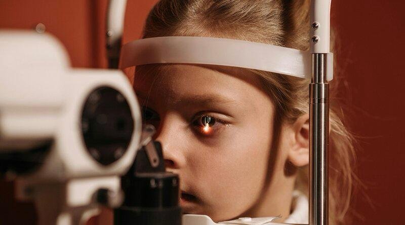
5. Diagnosis & Staging
Diagnostic work-up includes:
• Detailed ophthalmic examination under anaesthesia (fundoscopy, ultrasonography, optical coherence tomography)
• MRI of the brain and orbits to assess optic nerve and intracranial extension
• Classification systems: International Intraocular Retinoblastoma Classification (IIRC) and the newer AJCC 8th edition staging for retinoblastoma.
• Hereditary work-up (RB1 gene testing) for bilateral disease and family counselling
• Imaging (CT/MRI) and bone marrow / lumbar puncture if extra-ocular disease is suspected
6. Multidisciplinary Treatment Approach
A coordinated team including ocular oncologist, paediatric oncologist, interventional radiologist, radiation oncologist, geneticist, ophthalmic surgeon, and support services is essential.
Eye-salvage vs Enucleation
When possible, the goal is to preserve the eye and useful vision without compromising survival. Enucleation (removal of the eye) is considered when vision salvage is unlikely or tumour burden is extremely high (Group E eye in IIRC) or for extra-ocular disease.
Chemotherapy (systemic, intra-arterial, intravitreal)
• Systemic intravenous chemoreduction (VEC: vincristine, etoposide, carboplatin) to shrink tumour and minimise need for radiotherapy.
• Intra-arterial chemotherapy (IAC, ophthalmic artery infusion) has improved ocular salvage in advanced intra-ocular disease.
• Intravitreal chemotherapy (e.g., melphalan) for vitreous seeding — a major advance in preserving eyes with seeds.
Local therapies (laser photocoagulation, cryotherapy, brachytherapy)
After chemoreduction, focal treatments (laser, cryo, thermotherapy, plaque brachytherapy) eradicate residual tumour foci and seeds. Radiation therapy (external beam) is now reserved for specific high-risk cases due to long-term sequel risk of secondary malignancies.
Management of Metastatic/Extra-ocular Disease
Extra-ocular disease (optic nerve beyond lamina cribrosa, orbital extension, CNS metastases) requires intensive systemic chemotherapy, high-dose consolidation and stem cell rescue in some settings, plus radiotherapy. The prognosis is poorer but early multidisciplinary intensive care can achieve cure in selected cases.
7. Personalized & Precision Medicine in Retinoblastoma
Personalised care in retinoblastoma means:
• Tailoring treatment based on stage (intra-ocular vs extra-ocular), laterality (unilateral vs bilateral), tumour burden, seed status
• Genetic testing (RB1 status, MYCN amplification) influences surveillance and secondary-tumour risk
• Emerging biomarkers (liquid biopsy of aqueous humour) may guide future therapy decisions.
• Minimising long-term vision, cosmetic, endocrine and secondary malignancy risks by adjusting treatment modality (chemoreduction → local therapy rather than radiotherapy)
8. Follow-Up, Survivorship & Late Effects
Children treated for retinoblastoma require long-term follow-up for:
• Surveillance of second primary tumours (especially in hereditary cases)
• Vision monitoring and low-vision rehabilitation
• Psychosocial support, educational needs
• Screening for hearing, renal, cardiac toxicity (from chemotherapy)
• Cosmetic rehabilitation (prosthesis or ocular reconstruction if enucleation)
Thus, survivorship care is a key component of the multidisciplinary pathway.
9. How RecMed Supports Families Seeking Care Abroad
At RecMed Medical Travel (Istanbul, Türkiye), we coordinate:
• Rapid multidisciplinary review (ocular oncology, paediatric oncology, interventional radiology, genetic counselling)
• Access to advanced therapies: intra-arterial chemotherapy, intravitreal injections, focal therapies, genetic testing
• International patient logistics: visas, transport, accommodation, interpretation services
• Post-treatment coordination: long-term follow-up, vision rehabilitation, family counselling
By combining international referral expertise and personalised care pathways, we enable children with retinoblastoma to access best-in-class treatments while reducing delays and optimising outcomes.
10. Frequently Asked Questions (FAQ)
Is retinoblastoma curable?
Yes — in high-income countries the survival rate for intra-ocular retinoblastoma exceeds 95 % when diagnosed early and treated appropriately.
Will the child keep vision?
Vision salvage depends on tumour burden, seed status and treatment modality. The goal is eye and vision preservation when safe.
What is intra-arterial chemotherapy?
This involves delivering chemotherapy directly into the ophthalmic artery supplying the tumour‐bearing eye — enabling high local drug concentration while limiting systemic toxicity.
What is the risk of second cancers?
Children with heritable retinoblastoma (germline RB1 mutation) have increased risk of second malignancies later in life; lifelong surveillance is needed.
11. References
1. Ancona-Lezama D, et al. Modern treatment of retinoblastoma: a 2020 review. PMC. 2020.
2. Nag A., et al. Retinoblastoma – A comprehensive review, update and recent advances. PMC. 2024.
3. National Cancer Institute. Retinoblastoma Treatment (PDQ®). 2025.
4. Gupta AK, Meena JP. A narrative review of retinoblastoma and recent advances in its management. 2020.
5. Peng Z., et al. Global research landscape of retinoblastoma biomarkers. 2025.
6. Frontiers in Oncology. Treatment of Retinoblastoma: What Is the Latest and What’s Next? 2022.